PD-L1 – Immune Checkpoint
PD-L1 is the abbreviation for „Programmed cell Death Ligand 1“, a cell surface protein that is recognized by the receptor PD-1 (Programmed Death receptor 1) of certain immune cells. Some cancer cells display PD-L1 in high concentrations on their surface to prevent an attack by the immune system (a so-called immune evasion mechanism).[1] The clinical importance of PD-L1 arises from the fact that this evasion can be reverted by anti-PD-L1 monoclonal antibodies (mABs) such as durvalumab, atelizumab, or avelumab, or by anti-PD-1 mAbs such as nivolumab or pembrolizumab. The immune system is thus re-enabled to recognize and eradicate the cancer cells. This form of cancer immunotherapy is referred to as checkpoint inhibitor therapy (recognized with the Nobel Prize in Physiology or Medicine, awarded 2018 to the PD-1 discoverer Tasuku Honjo).[2]
Since PD-L1 is only expressed by a fraction of cancers, agents for clinical imaging of PD-L1 are of high interest to select those patients that are likely to benefit from said immunotherapies,[3] or to monitor the progress of ongoing therapies.[4]
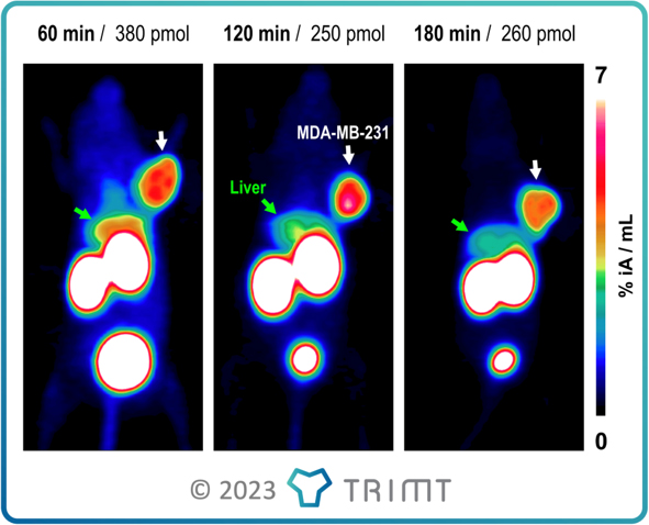
TRIMT’s portfolio includes tracers for positron emission tomography (PET) imaging of PD-L1, labeled with the short-lived medical isotope Gallium-68 (68Ga). Preclinical imaging confirmed favorable properties for sensitive imaging of PD-L1 expression by PET (see Image).[5]
[1] Ribas A, Wolchok JD. Science 2018;359:1350.
[2] https://en.wikipedia.org/wiki/Tasuku_Honjo
[3] Krutzek F, et al., Pharmaceuticals 2022;15:747.
[4] Nimmagadda S, Cancers 2020;12:3173.
[5] Notni J., et al., unpublished results.